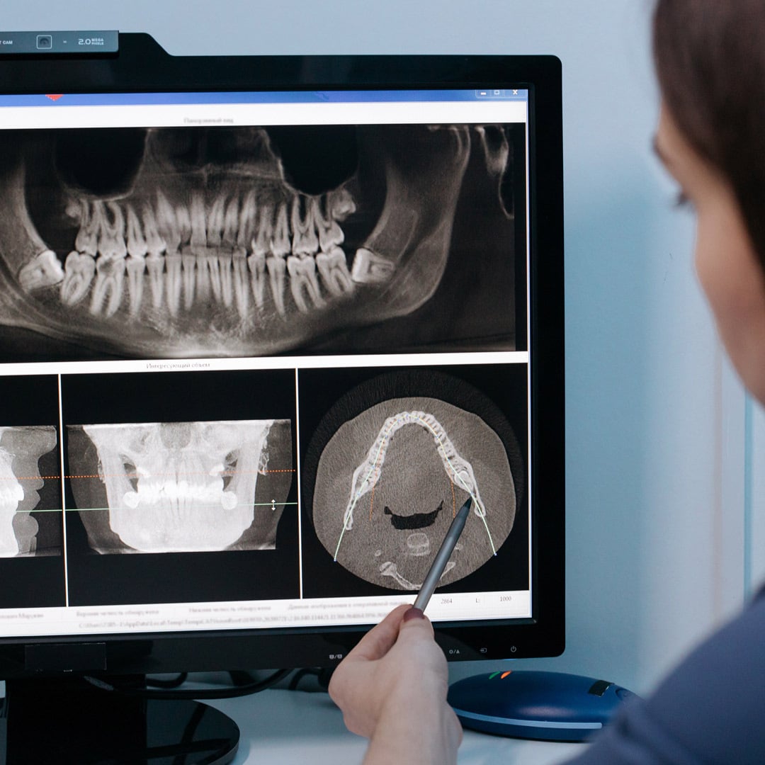
You are probably used to a dental technician taking x-rays of your teeth at your regular dental checkups. This is an important part of the diagnostic process, which allows your dentist to evaluate the health of your teeth, bones and soft tissues. These images are most commonly used to spot cavities, but they also can detect bone loss, root damage and hidden dental structures–such as impacted teeth.
What are X-Rays?
Dental images are created using x-rays, a high-energy electromagnetic radiation that can permeate or be absorbed by solid objects. Dense objects, such as teeth and bones, that absorb the energy appear as light-colored areas in the image. Less dense objects, such as gums and cheeks, produce dark areas on x-ray film.
Because so much of your dental health lies below the surface, x-rays allow your dentist to detect problems that an oral exam won’t show. X-rays can help the dentist identify newly developing cavities or areas that have the potential to become cavities. X-rays also can detect cavities in areas between the teeth that would be difficult to discover with a physical exam.
X-rays allow the dentist to observe changes in the root canal, between the gum and a tooth, or within a supporting bone that could result from an infection. In children and teenagers, x-rays can help determine whether the mouth contains enough room for all developing teeth or whether wisdom teeth are impacted.
Types of X-Rays
Two main classifications of dental x-rays exist: intraoral and extraoral. Intraoral x-rays are captured from inside the mouth, while extraoral x-rays are captured from outside.
Intraoral x-rays are the most commonly used. They focus on the teeth and provide crisp detail. When taking these pictures, the technician will ask you to bite a small device used to capture the images. The most common types of intraoral x-rays are bitewing, periapical and occlusal.
- Bitewing x-rays show the position of the upper and lower teeth. The images reveal the health of the enamel, inner canals and roots. Enamel and fillings are dense and appear white in color, whereas the bones surrounding areas of decay are less dense and therefore appear darker. Orthodontists are trained to interpret the light and dark patterns to distinguish healthy teeth from damaged ones.
- Periapical x-rays show the whole tooth–from crown to root–in one portion of the jaw. Dentists use them to detect changes in the root and surrounding bone structures. Each periapical x-ray shows a small section of your upper or lower teeth.
- Occlusal x-rays track the development and placement of an entire arch of teeth in either the upper or lower jaw. Occlusal x-rays show the roof or floor of the mouth and are used to find extra teeth, teeth that have not yet broken through the gums, jaw fractures, a cleft palate, cysts, abscesses or growths.
Extraoral x-rays focus mainly on how the jaw’s positioned in relation to the teeth. These x-rays are not as detailed as intraoral ones and are not meant to diagnose problems within specific teeth. They also are free of any discomfort because the imaging is done from outside of the mouth without interaction from the patient–aside from holding still. The most common types of extraoral x-ray are panoramic, cephalometric, and tomographic.
- Panoramic x-rays display the entire mouth in a single image. This includes the teeth, upper and lower jaws, tissues and surrounding structures. An orthodontist often uses a panoramic x-ray to assess gum and bone irregularities that will help him plan for treatment.
- Cephalometric x-rays capture an image of the side of the head and illustrate the relationship between the teeth, jaw and profile. This type of x-ray is especially helpful when planning orthodontic treatment because it will expose misalignments that treatment can correct.
- Cone-beam computed tomography (CBCT) creates 3-D images of the teeth and mouth using a cone-shaped x-ray beam. Tomographic x-rays are useful for examining structures that are difficult to see clearly because of an obstruction.
Are X-Rays Safe?
Some people wonder if x-rays are safe since they require low levels of radiation exposure–less than you would receive when being screened for an airline flight. While the health risks for a single dental x-ray is very small, dentists take every possible precaution to limit unnecessary exposure.
According to the American Dental Association, “How often x-rays should be taken depends on your present oral health, your age, your risk for disease, and any signs and symptoms of oral disease. Dentists take precautions to ensure that the low level of radiation exposure—both to patients and to dental professionals—is ‘As Low As Reasonably Achievable’ (the ALARA principle).”1
If you are switching between providers or specialists, you can request copies of your dental records, including recent x-rays, from your previous provider to prevent duplicate procedures. If you are pregnant or have pre-existing conditions, discuss with your dentist any concerns you may have about dental imaging.
How Often Should I Get Dental X-Rays
The frequency of dental imaging can vary greatly between patients. Typically, a new patient will receive a full-mouth x-ray workup at the initial visit and then every three years subsequently.
Many guidelines suggest that bitewing x-rays should be done at least once a year and twice a year for patients considered at a higher risk due to conditions such as dry mouth, an abundance of fillings or crowns, or periodontal disease. These patients might require full x-rays more frequently as well. Additionally, children and adolescents will generally need to receive x-rays more frequently than adults.
X-Rays and Braces
Dental imaging is an important part of your orthodontic treatment before, during and after braces. The types of x-ray the orthodontist will need depends on your treatment plan. Initial x-rays identify problems that need to be corrected before braces. They also allow the orthodontist to consider the health and placement of the roots, soft tissues, muscles, joints, nerves and bone structures that all will factor into your treatment plan.
Some things your orthodontist will look for include
- misshaped or misplaced teeth or roots,
- abnormal length of teeth or roots,
- jawbones that are misaligned, asymmetrical, incorrectly sized or improperly spaced.
X-rays at different stages in your orthodontic treatment will monitor how your realignment is progressing and how your roots and jaw are responding to the shift plan.
A final set of x-rays once your braces are removed will allow the orthodontist to evaluate the outcome of treatment and make recommendations for other procedures if needed. He will also use these images to create your retainer. You will most likely wear your retainer every day and night for several months.
What Types of Imaging Do Orthodontists Use?
Orthodontists now use both intraoral and extraoral imaging technology to create impressions of your teeth without having to create bite impressions from a plaster mold. The digital images the scanners create enable orthodontists to pinpoint small structural problems and plan more effective treatment compared to the traditional molds.
3M True Definition Scanner
True Definition scanners can download and save hundreds of impressions digitally, resulting in greater comfort and convenience for the patient and increased precision for orthodontists and their staff. They allow the orthodontist to take impressions of the upper and lower sets of teeth without the patient having to bite, twist, turn or otherwise manipulate the process. These images will not degrade over time, which is important when comparing progress from early to later phases of treatment.
iTero Scanner
Another intraoral imaging technology eliminating the need for impressions is the iTero scanner. Like the True Definition, the iTero is a hand-held wand. It uses a radiation-free laser that the technician runs over your teeth, creating a 3D digital impression of your teeth and soft tissue structures within minutes. Through digital software, your orthodontist can follow the progression of the scans over time to ensure treatment is proceeding according to plan.
Cone-Beam Computed Tomography
The CBCT is an extraoral X-ray that creates a 3D computer model of your teeth and helps your treatment provider analyze the orientation of your teeth and their roots. It also allows for your treatment provider to perform more complex procedures and correct more serious problems.
Whether digital or x-ray technologies are used, dental imaging is an important part of your orthodontic treatment. Schedule a consultation with one of our board-certified orthodontists today.


